Health + Medicine
Feature
Repairing Defects Before Birth

It’s about as far from a conventional doctor-patient relationship as you get. The doctors not only need no bedside manner, ideally they will never talk to their patients or see them as more than images on a monitor. And the patients will never describe symptoms to their doctor or voice complaints.
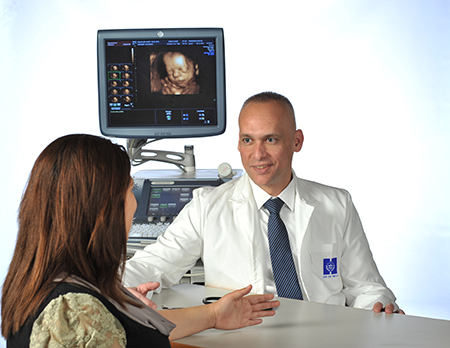
These patients are fetuses and their doctors, fetal medicine specialists. “Until recently, maternal and fetal health was a single entity, but the new specialty separates them,” says Dr. Yuval Gielchinsky, a senior physician in the Department of Obstetrics and Gynecology at the Hadassah–Hebrew University Medical Center at Ein Kerem. “Fetal medicine assesses the well-being of the developing child from eight weeks after conception, at the end of the embryo stage, until birth. It diagnoses fetal abnormalities and illnesses and treats them.”
A Hadassah-trained obstetrician with a Ph.D. in molecular biology, Dr. Gielchinsky established Hadassah’s Center for Fetal Medicine five years ago, following a fellowship at King’s College London with fetal medicine trailblazer Dr. Kypros Nicolaides. Hadassah’s center is one of only a handful worldwide, a multidisciplinary unit that is leading the way in Israel to better fetal care.
“Medical science has been able to diagnose problems in the fetus for some time,” says Dr. Gielchinsky. “It tested amniotic fluid for Down syndrome and looked for physical abnormalities on blurry ultrasounds. Today, with high-resolution ultrasound, more advanced imaging technologies and better understanding of what to look for, we’re able not only to identify several hundred chromosomal and genetic disorders, but also to measure blood flow and check whether the developing fetus is anemic.”
Even better, according to Dr. Gielchinsky, are the numerous ways already developed—and in development—to treat these conditions. “The majority of pregnancies are problem-free,” he says. “But in those that aren’t, there are interventions.”
“Katya” is one Hadassah patient who delivered a healthy infant after noninvasive treatment. “During a routine scan early in my pregnancy, they picked up an irregular heartbeat in my baby,” she explains. “I took medication to control his abnormal heart rhythms, a medication that passed into him through the placenta. The dose was too small to affect me, but it was enough to save his life. He was born at term, whole and perfect.”
The Doppler fetal monitor, long used to measure heartbeat, can also gauge the flow of blood through the fetus—and give timely warning if anything is amiss. “Overly rapid blood flow indicates that the blood is too dilute,” says Dr. Gielchinsky. “One likely reason is that the blood is low in hemoglobin. From its speed of flow, we can calculate the hemoglobin level and thus determine whether the fetus is anemic.”
Fetal anemia can result from an infection in the mother (Parvovirus is one well-known culprit) or when the fetus inherits from its father a type of red blood cell protein that the mother lacks—the Rhesus or Rh factor. The Rh immunoglobulin shot is preventive only; in women already sensitized to the Rh factor, it is not effective, and the mother’s immune system misidentifies this protein as an intruder, creating antibodies to destroy it and, along with it, the fetal red blood cells.
“Red blood cells transport oxygen to all the body’s cells and organs,” says Dr. Gielchinsky. “If they’re inadequate, the fetal heart compensates by pumping harder, which can lead to heart failure, even brain damage. Now we can treat an anemic fetus by transfusing blood through the mother’s abdominal wall into the umbilical cord.”
If transfusing a fetus sounds futuristic, it pales in comparison with surgery inside the womb. The main surgical tool is the fetoscope—a laparoscope just millimeters thick with a fiber-optic camera and channels to insert instruments. It is slid into the uterus from a small incision in the mother’s abdomen, its camera transmitting images to the physicians.
“Its most common clinical use is in a condition that affects identical twins,” says Dr. Gielchinsky. “In about one in five such pairs, the blood vessels in the shared placenta are abnormally routed and connect the circulations of the two fetuses.”
Known as twin-to-twin transfusion syndrome, this unique complication of monochorionic twins—twins sharing a placenta—directs volumes of blood from one fetus into the other. The “donor” twin, starved of blood, is small and less developed. The “recipient” twin is overloaded and will likely suffer congestive heart failure, trying to pump vast amounts of blood through its system.
“We disconnect them with a technique developed by Dr. Nicolaides,” says Dr. Gielchinsky. “We locate the shared blood vessels on images transmitted by the fetoscope’s camera and seal them with a laser. Each twin is left with its own link to the placenta through which it receives its own share of nutrients.”
Performed only when the twin fetuses are at great risk, Hadassah’s results are above average: 82 percent of procedures result in one live baby, and 60 percent in two live babies.
Another life-saving procedure performed at Hadassah guides a tiny balloon through a fetus’s mouth and into the windpipe. This is an out-of-the-box treatment for congenital diaphragmatic hernia, a condition known as CDH, affecting about one in every 2,500 babies.
“CDH is a hole in the diaphragm,” explains Dr. Gielchinsky. “Stomach, spleen, liver and intestines drift up through this hole, filling the chest cavity. It’s usually repaired surgically after birth, but some affected babies won’t survive birth because their lungs are insufficiently developed. We plug the trachea or windpipe by inflating a balloon inside it—popping and removing it a few weeks later. Fluid is thus trapped inside the lungs, inflating them and staking space in which they can grow.”
CDH and twin-to-twin transfusion syndrome are life-threatening conditions. Spina bifida, a congenital disorder that occurs in one in 1,500 pregnancies, does not kill, but its impact on quality of life is devastating. When the spine fails to fuse around the spinal cord, leaving nerves exposed, infants are usually born with paralyzed lower limbs, no bladder or bowel control and, because of cerebrospinal fluid pooling in the brain, mental disabilities. Usually picked up at the 20-week scan, over 60 percent of couples choose to terminate a spina bifida pregnancy.
“Closing the spinal cord in utero can’t reverse nerve damage, but will prevent further injury and accumulation of fluid in the brain,” says Dr. Mony Benifla, head of Hadassah Hospital’s Department of Pediatric Neurosurgery.
One way of repairing the spinal cord before birth is by opening the uterus and amniotic sac, partially extracting the fetus (kept facedown in amniotic fluid), pushing the nerve bundle back inside the vertebrae, patching the wound, replacing the fetus and closing the mother’s uterus and abdomen.
The other way is surgically more complex but far less traumatic to mother and fetus: It is keyhole surgery, performed through three fetoscopes inserted into the uterus.
“We’ve been learning the technique and will introduce it to Hadassah and Israel,” says Dr. Benifla. “Dr. Gielchinsky will get us to the fetus and its exposed spine, and I will perform the repair. It’s far from easy, but by preventing exposure of the nerves to amniotic fluid in utero and to the outer world after delivery, it gives the child a better chance of a normal IQ, better bowel and bladder control and better lower limb movement.”
With anesthesiologist Dr. Yehuda Ginosar, Drs. Benifla and Gielchinsky have traveled to São Paulo to train with one of the procedure’s pioneers, Dr. Denise Pedreira, at the Hospital Israelita Albert Einstein. São Paulo’s Jewish community established the hospital, and Hadassah International’s Brazil branch has nurtured this partnership. “Denise Pedreira has performed the operation dozens of times since bringing it to Brazil in 2013,” says Dr. Benifla. “She’ll come to Hadassah to help us launch—making Israel one of the first countries outside Brazil where it’s offered.”
Fetal medicine, like pediatric and adult medicine, already has subspecialties. Its multidisciplinary team includes anesthesiologists, neonatologists, pediatric surgeons and operating room nurses. Among key members of Hadassah’s team are Dr. Ginosar, director of Hadassah’s Mother and Child Anesthesia Unit, who performs almost all fetal anesthetics; Dr. Smadar Eventov-Friedman, head of the Neonatal Intensive Care Unit; and operating room nurses Orna Ben-Yosef and Marina Bineashvilli.
Fetal medicine is a highly skilled multidisciplinary specialty, and Hadassah currently has staff and facilities for only one major intervention every two weeks. With a fetus increasingly treated as independent of its mother, however, fetal medicine—at Hadassah and elsewhere—will only expand.
Wendy Elliman is a British-born science writer who has lived in Israel for more than four decades.


 Facebook
 Instagram
 Twitter
[…] Schäden schon vor der Geburt korrigieren Chirurgen im Jerusalemer Hadassah-Krankenhaus können Anomalien im Fötus ab dem Alter von acht Wochen nach der Empfängnis aufspüren und korrigieren — unregelmäßiger Herzschlag, Anämie, Zwillinge, die sich eine Plazenta teilen, angeborene Hernie, Korrektur des Rückenmarks. Israelische Ärzte retten Leben, noch bevor sie begonnen haben! http://www.hadassahmagazine.org/2016/12/22/repairing-defects-birth/ […]